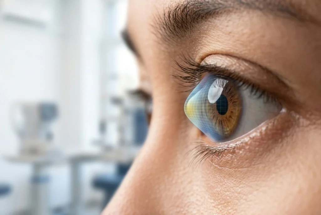
Keratoconus is a condition in which the cornea gradually becomes thinner and changes shape, becoming more protruded or cone-shaped. Changes in the shape of the cornea can prevent light from focusing properly, resulting in blurred, shadowed, or distorted vision.
Keratoconus usually develops gradually and can affect both eyes, although the severity may differ between the two eyes.
What Is Keratoconus?
Keratoconus is a progressive disorder of the cornea that causes changes in its shape and strength. This condition can lead to irregular astigmatism, blurred vision, and reduced visual quality.
Early detection is important because timely treatment can help maintain the condition of the cornea and preserve visual function.
What Is Keratoconus?
Keratoconus is a corneal disorder in which the cornea gradually becomes thinner and protrudes forward.
Changes in the shape of the cornea can interfere with the way light is focused onto the retina. As a result, vision may become unclear even when a person is wearing glasses. Keratoconus generally begins at a young age and may continue to change over time.
What Are the Symptoms of Keratoconus?
Symptoms of keratoconus may include blurred vision, changing astigmatism, and shadowed or distorted vision.
Some symptoms that may occur include:
- Increasingly blurred vision.
- Frequent changes in astigmatism prescription.
- Shadowed or ghost images.
- Straight lines appearing distorted or wavy.
- Difficulty seeing clearly even when wearing glasses.
- Increased sensitivity to light.
- Frequently squinting to see more clearly.
- Increasing difficulty seeing at night.
In its early stages, the symptoms may resemble ordinary nearsightedness or astigmatism.
Is Keratoconus the Same as Astigmatism?
No, they are not the same.
Astigmatism is a refractive error that occurs when the surface of the cornea or lens has an uneven curvature. In keratoconus, changes in the shape of the cornea can progressively worsen and result in more irregular astigmatism.
Therefore, high astigmatism, rapidly changing astigmatism, or astigmatism that is difficult to correct with glasses may be a reason to undergo further corneal evaluation.
What Causes Keratoconus?
The exact cause of keratoconus is not fully understood. The condition is thought to involve a combination of genetic and environmental factors.
Some factors that may be associated with keratoconus include:
- A family history of keratoconus.
- Excessive eye rubbing.
- Allergies or certain conditions that cause frequent eye itching.
- Certain genetic factors.
Having one of these factors does not necessarily mean that a person will develop keratoconus.
Can Eye Rubbing Cause Keratoconus?
Excessive eye rubbing may be associated with keratoconus and its progression.
Eye rubbing places mechanical pressure on the cornea. In individuals with certain corneal susceptibility, this habit may contribute to worsening of the condition. Therefore, excessive eye rubbing should be avoided, particularly in people who already have a corneal disorder.
How Is Keratoconus Detected?
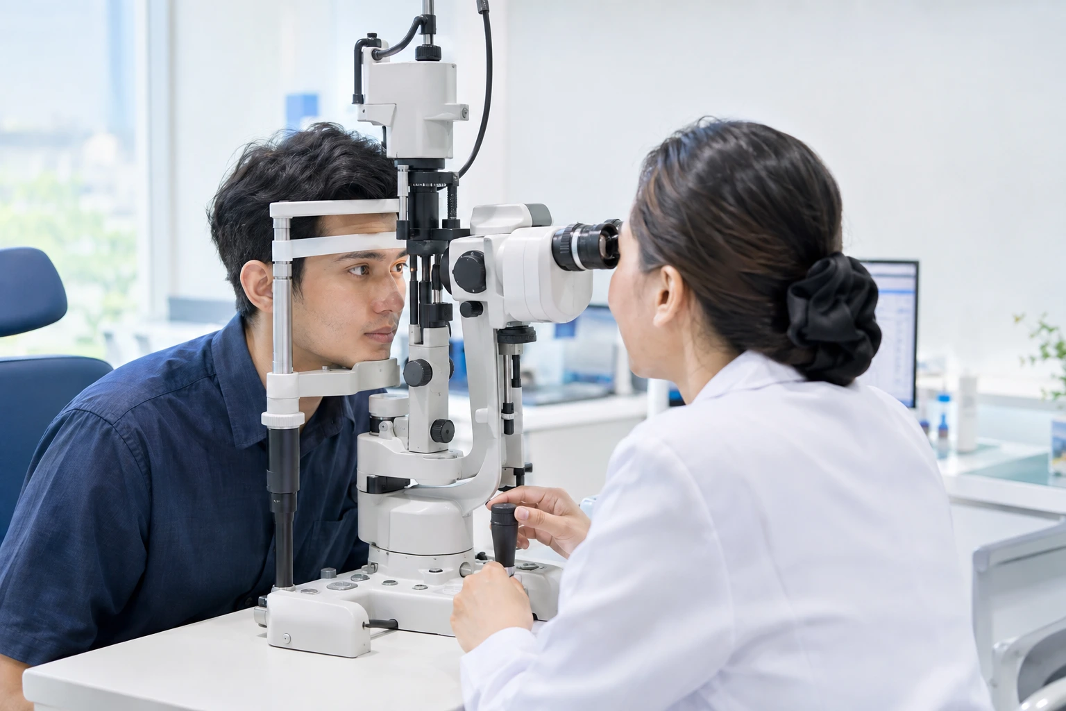
Keratoconus cannot be diagnosed based solely on a nearsightedness or astigmatism test. An examination of the shape and structure of the cornea is necessary.
Doctors may perform examinations such as:
- Visual acuity testing.
- Refraction testing.
- Corneal topography.
- Corneal tomography.
- Corneal thickness measurement.
- Corneal biomechanical assessment, when necessary.
These examinations help evaluate the shape, thickness, and characteristics of the cornea, allowing changes associated with keratoconus to be detected.
What Are Corneal Topography and Tomography?
Corneal topography and tomography are imaging examinations used to evaluate the shape of the cornea. Topography provides a map of the corneal surface.
Tomography provides more comprehensive information about the shape and structure of the cornea, including its front and back surfaces as well as other parameters. These examinations help doctors determine whether corneal changes are consistent with keratoconus or another corneal condition.
Can Keratoconus Be Cured?
Keratoconus can be treated, but the changes in corneal shape that have already occurred cannot always be completely reversed. The goals of treatment are to help maintain the condition of the cornea, improve visual quality, and, in certain cases, slow down or stop disease progression. Treatment options depend on the severity of keratoconus and each patient’s individual condition.
How Is Keratoconus Treated?
Treatment may vary depending on the stage of the disease.
- Glasses
In the early stages of keratoconus, glasses may help improve vision. However, as the shape of the cornea changes, glasses may no longer provide sufficiently clear vision. - Specialized Contact Lenses
In certain cases, doctors may recommend specialized contact lenses to help improve visual quality. The type of lens is selected based on the patient’s corneal shape and condition. - Corneal Cross-Linking
Corneal cross-linking (CXL) is intended to strengthen the corneal tissue and help slow the progression of keratoconus. This procedure is one of the treatment options for certain patients, particularly when examinations indicate that the keratoconus is still progressing.
For more specific information about this procedure, you can read the article Cross-Linking: A Modern Treatment for Keratoconus.
Corneal Procedures or Surgery
In advanced keratoconus that cannot be adequately corrected with the options above, doctors may consider certain corneal procedures, including surgical treatment in cases where it is indicated. The choice of treatment depends greatly on the condition of the cornea and the patient’s visual quality.
Can Keratoconus Cause Blindness?
Keratoconus can cause significant vision loss, but it does not always result in complete blindness. As keratoconus progresses, changes in the shape and clarity of the cornea may increasingly affect visual quality. Therefore, early detection and regular monitoring are important to determine whether the condition is progressing.
Can Keratoconus Be Treated with LASIK?
LASIK is generally not recommended for patients with keratoconus.
Before undergoing a laser procedure such as LASIK, the condition and shape of the cornea must be carefully evaluated. In keratoconus, the corneal structure has already undergone changes, and procedures that remove corneal tissue may increase the risk of corneal problems in certain patients. Therefore, patients with nearsightedness or astigmatism who are considering LASIK should undergo a corneal evaluation beforehand.
Can Keratoconus Be Prevented?
Not all cases of keratoconus can be prevented because genetic factors may also play a role. However, certain habits may help maintain corneal health, particularly avoiding excessive eye rubbing.
If someone has a family history of keratoconus or experiences rapid changes in astigmatism prescription, a corneal examination can help detect changes at an earlier stage.
When Should You Have an Eye Examination?
An eye and corneal examination should be considered if you:
- Experience frequent changes in astigmatism prescription.
- Have increasingly blurred vision even when wearing glasses.
- Experience shadowed or distorted vision.
- Have increasing difficulty seeing at night.
- Frequently rub your eyes.
- Have a family member with keratoconus.
- Have previously been suspected by a doctor of having a corneal disorder.
The earlier corneal changes are detected, the sooner a doctor can determine whether the condition only needs monitoring or requires treatment.
FAQ About Keratoconus
Keratoconus is a progressive disorder of the cornea that causes the cornea to become thinner and change shape, which can result in blurred or distorted vision.
Symptoms may include blurred vision, frequently changing astigmatism, shadowed vision, and difficulty achieving clear vision even when wearing glasses.
No. High astigmatism does not automatically mean that someone has keratoconus. However, high, irregular, or frequently changing astigmatism may be a reason to undergo further corneal evaluation.
Keratoconus can be treated to help maintain the condition of the cornea and visual function. Treatment options depend on the severity and progression of the disease.
LASIK is generally not recommended for patients with keratoconus because the condition of the cornea requires special consideration. A corneal examination is necessary before determining the appropriate vision correction option.
Keratoconus can cause significant vision loss if it progresses, but it does not always result in complete blindness. Early detection and monitoring are important for managing the condition.
Conclusion
Keratoconus is a corneal disorder that causes the cornea to gradually become thinner and change shape. This condition can result in irregular astigmatism, blurred or shadowed vision, and reduced visual quality.
The early symptoms of keratoconus may resemble ordinary nearsightedness or astigmatism. Therefore, corneal topography or tomography is important when there are rapid changes in refractive errors or a suspected corneal disorder.
Treatment may include glasses, specialized contact lenses, corneal cross-linking, and certain corneal procedures, depending on the patient’s condition.
If your vision is becoming increasingly blurred or your astigmatism prescription changes frequently, an examination by an ophthalmologist can help detect keratoconus and determine the appropriate treatment.
Reference:
National Eye Institute – Keratoconus
American Academy of Ophthalmology – Keratoconus
American Academy of Ophthalmology EyeWiki – Keratoconus